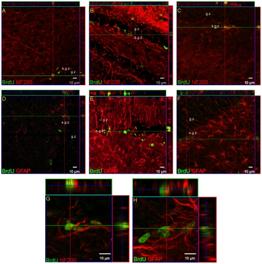
Figure 2. Newly-divided cells in the DG of adult gerbil hippocampus after global ischemia.
Animals were subjected to 5 min of global forebrain ischemia followed by reperfusion. BrdU was administered 24 h prior to sacrifice. Brain sections were double-labeled with anti BrdU antibody (green) and anti-NF-200 (red) (A, B, C, G) or anti-GFAP (red) (D, E, F, H). Confocal photomicrographs show immunohistochemical reaction in control DG (A, D), 9 days after ischemia (B, E), and 28 days after ischemia (C, F). G, H represent magnification (z-stacks) of the picture C and F. Photomicrographs are representative of observations made from six animals per time point. Scale bar 10 µm. Abbreviations: s.g.z – subgranular zone, g.z. –granular zone.

