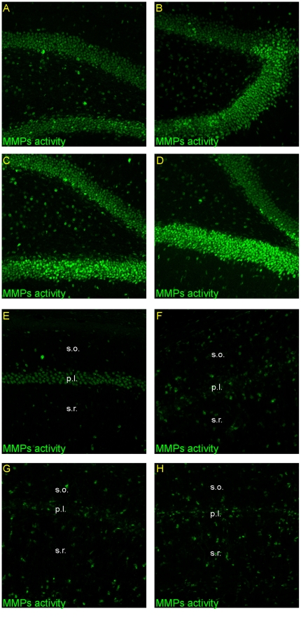
Figure 5. Activity of metalloproteinases in the adult gerbil hippocampus after global ischemia.
Animals were subjected to 5 min of global forebrain ischemia followed by reperfusion. Confocal photomicrographs showing in situ zymography in DG and CA1 from a control animal (A, E) and from ischemic animals sacrificed at 7 (B, F), 14 (C, G) and 28 days (D, H) after ischemia. Note the increase of fluorescence signal in the DG and in strata oriens and stratum radiatum with simultaneous decrease in pyramidal cell layer of the CA1 area across the time of reperfusion. All images subjected to direct comparisons were captured at the same exposure and digital gain settings. Photomicrographs are representative of observations made from six animals per time point. Abbreviations: s.o – stratum oriens; s.r. – stratum radiatum.

