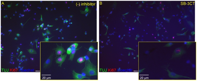
Figure 9. Effect of SB-3CT (10 µM) on the Ki67 and Tuj1 positive cells in HUCB-NSs culture.
Equivalent numbers of HUCB-NSCs were plated on fibronectin-coated coverslips and grown 8 days in serum-free medium with or without SB-3CT (10 µM). Cells were stained for Ki67 (red) and Tuj1 (green). The cell nuclei counterstained with Hoechst (blue). Note the decreased number of immunolabeled cells in the presence of SB-3CT. Scale bar 20 µm.

