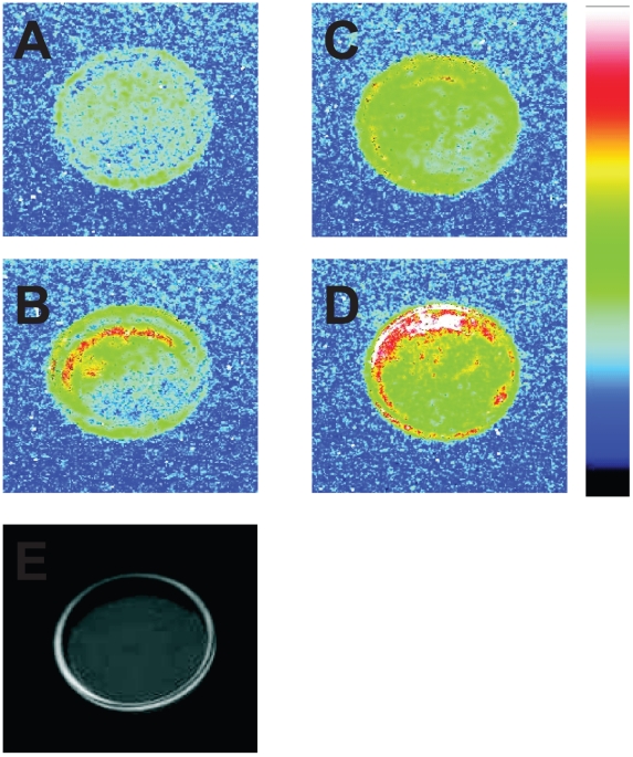
Figure 1. Two-dimensional imaging of the ultra-weak photon emission from the intact (A, C) and the disrupted (B, D) cells measured in the absence (A, B) and the presence (C, D) of linoleic acid.
Prior to the measurements, the cells suspended in Tris-Acetate Phosphate buffer (pH 7.2) were placed on the Petri dish and kept for 30 min in the dark. In (B, D), prior to the dark adaptation the cells were frozen in liquid nitrogen and subsequently warmed to room temperature. In (C, D), 2 mM linoleic acid was added to the cells prior to the measurements. (E) represents photograph of the cells placed on the Petri dish taken under weak light illumination. Ultra-weak photon emission imaging was measured using a highly sensitive CCD camera with an integration time of 30 min.

