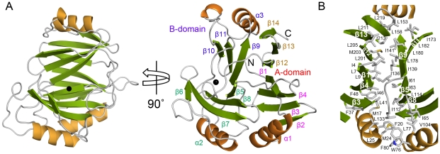
Figure 2. Overall structure of TxDE in the substrate-free form.
A, A ribbon diagram is shown, with the corresponding secondary structures labeled as defined in Figure 1B. The black sphere indicates the Mn(II) ion. In the left panel, the molecule is oriented to place the B-domain in front, and a different view is shown in the right panel. Labels for the secondary structures in each motif are indicated in different colors. B, Hydrophobic interactions between the A- and B-domains are shown. Hydrophobic residues such as leucine, isoleucine, and phenylalanine are predominantly located in this interdomain interface. These residues along the β-sheet include Ile-213, Leu-211, Leu-205, Met-203, Leu-201, Ile-4, Leu-7, Leu-9, Ile-46, Phe-48, and Leu-41 from the A-domain, and Ile-173, Leu-178, Leu-180, Leu-182, Ile-136, Ile-139, Ile-141, Ile-61, Ile-63, and Ile-112 from the B-domain.

