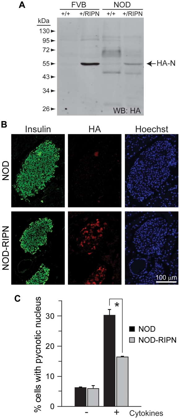
Figure 1. Expression levels and functionality of the RIPN transgene in NOD mice.
A. Western blot analysis of lysates from islets isolated from ten week-old female mice. The presence of fragment N was assessed using an anti-HA antibody. B. Immunohistochemistry analysis of paraffin sections of five week-old female mice. Sections were stained using anti-insulin and anti-HA antibodies. Nuclei were stained with Hoechst 33342. C. Freshly isolated islets from 5 week-old females were incubated or not with inflammatory cytokines (1,000 units/ml TNFα, 1,000 units/ml interleukin-1β, and 50 units/ml interferon-γ) during 24 hours. The islets were then stained with Hoechst 33342 and apoptosis was scored.

