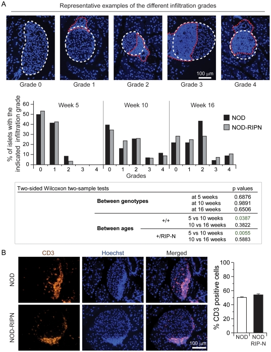
Figure 3. Lymphocytic infiltration.
A. Paraffin sections of female mice of the indicated ages were stained with Hoechst 33342. Infiltration was scored as described in the methods. The upper images depict representative examples of the different infiltration grades (the white dotted lines delineate the islets and the red dotted lines encircle the lymphocytic infiltration). B. Paraffin sections of infiltration grade 2 islets of 10 weeks old NOD and NOD-RIPN mice were stained with an anti-CD3 specific antibody (orange). The nuclei were stained with Hoechst 33342 (blue). Quantitation of CD3-positive cells relative to the total number of islet cells is shown on the right. Note that T cells (i.e. CD3-positive cells) are smaller than islet cells. Hence T cells making about 50% of the total number of cells within islets occupy an islet area that is less than half.

