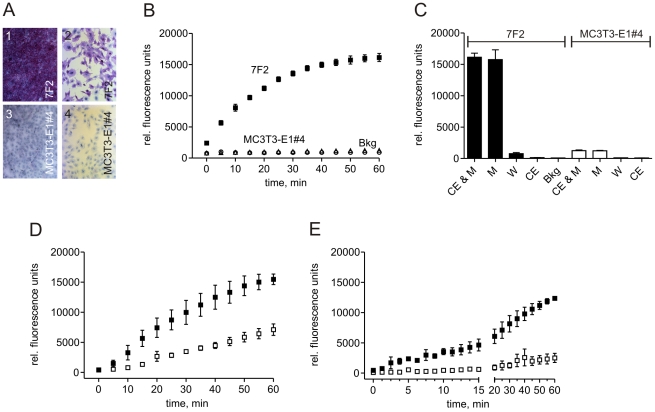
Figure 2. ALP-dependent DiFMUP activation on the surface of ALP-positive osteoblastic bone cells and in the presence of rat tibial bone.
A: Histochemistry detected ALP expression as violet-red staining on 7F2 cells as shown in low and high magnification images 1 and 2, respectively. In contrast, no ALP expression was detectable on the MC3T3-E1#4 cells shown also in low (3) and high magnification (4). B: Activation (hydrolysis) of DiFMUP occurred on 7F2 cells (closed squares), but not on ALP-negative MC3T3-E1#4 cells (open triangles). No-cell background (Bkg) measurements are also presented (open circles). C: Following the separation of cells (CE) and medium (M) and a single wash step (W), the vast majority of the hydrolysis product was found in the medium. Mean values and standard deviations are shown (n = 3). D: DiFMUP in physiological solution was activated in a time-dependent fashion in the presence of highly purified rat tibia bone (closed squares). Addition of the ALP inhibitor levamisole reduced DiFMUP activation (open squares). E: DiFMUP in alkaline solution was activated by a single highly purified tibia bone chip (closed squares). Presence of levamisole significantly suppressed the activation (open squares). Mean values and standard deviations are shown (n = 3).

