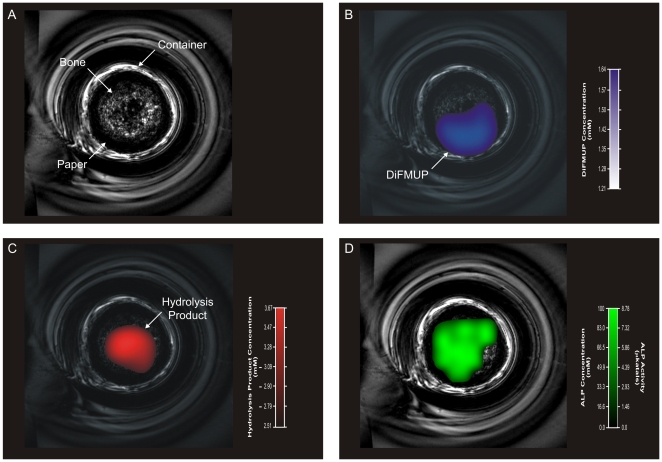
Figure 4. Non-invasive imaging of ALP activity in rat tibia cortical bone.
A: RARE 1H images of the bone sample anatomy including the rat tibia cortical bone core within the glass vial. B and C: 19FMRSI-derived parametric maps of regional DiFMUP and hydrolysis product concentrations overlaid onto RARE 1H images of the bone sample (B and C, respectively). D: 19FMRSI-derived parametric maps of regional ALP concentration and activity overlaid onto RARE 1H images of the bone sample.

