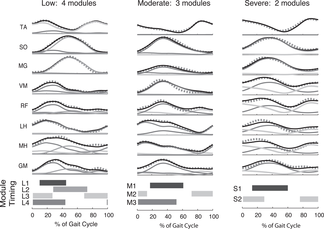
Figure 3.
Decreased complexity of locomotor muscle coordination patterns in hemiparetic stroke subjects. As the level of gait impairment worsens (as determined by walking speed and gait asymmetry), muscle activation patterns in the affected limb become more abnormal (black lines). Muscle synergy analysis reveals that fewer muscle synergies (modules) are needed to reconstruct the muscle activity during locomotion. Thin solid lines indicate individual contributions of muscle synergies, and thick dotted lines indicate muscle synergy reconstructions. Horizontal bars indicate where in the gait cycle each muscle synergy is most actively recruited (defined as >50% of mean activity). When 4 muscle synergies are available, they are activated in a phasic pattern typical of normal walking (left column, shaded bars). However, as the number of muscle synergies decreases, they become active for a greater proportion of the gait cycle (middle and right column). TA = tibialis anterior; SO = soleus; MG = medial gastrocnemius; VM = vastus medialis; RF = rectus femoris; MH = medial hamstrings; LH = lateral hamstrings; GM = gluteus medius. Adapted, with permission, from Clark DJ, et al. Merging of healthy motor modules predicts reduced locomotor performance and muscle J Neurophysiol. 2010;103(2):844–857. Copyright 2010 by the American Physiological Society.

