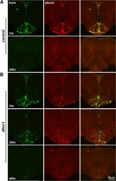
Figure 9. Clearance of bursicon neurons is likewise delayed in the dfmr1 null brain.
Representative images of the ventral brain SEG double-labeled for bursicon (green) and pburs (red) in control (w1118) and dfmr150M null mutants. A) A control brain at 1 hour post-elcosion (top row) shows the two bursicon peptidergic neurons' somal positioning and process elaboration. The bottom row reveals that the neurons have been eliminated by 24 hours post-eclosion in the wild-type brain. B) Comparable images from the dfmr1 null brain at 1 hour post-elcosion (top row) show a complex neuronal array indistinguishable from control. Bursicon processes are aberrantly retained at 24 hours in the dfmr1 null mutant (middle row), although largely lost from the brain SEG by 48 hours (bottom row).

