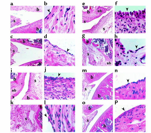
Figure 8.
Inhibition of ongoing AA by MCP-1–targeted DNA vaccination modulates histological changes in the acute and chronic phase of disease. Thirty and 90 days after AA induction joint samples from each of the experimental groups described in Figure 7a (four representative rats per group) were subjected for histological analysis (12 sections each group). (a, c, e, g, i, k, m, and o) Representative synovial joints (×5). (b, d, f, h, j, l, n, and p) Representative synovial tissue (×40). a and b show naive joints taken, with age matching to the experiment rats (day 30); c and d show naive joints taken, with age matching to the experiments rats (day 90). e and f show arthritic joints taken 30 days after disease induction, and g and h arthritic joints taken 90 days after disease induction. i and j show pcDNA3-treated joints taken 30 days after disease induction, k and l show pcDNA3-treated joints taken 90 days after disease induction. m and n show MCP-1–treated joints taken 30 days after disease induction, o and p show pcDNA3-treated joints taken 90 days after disease induction. The arrowheads point to the synovial lining. b, bone; nb, new bone formation; s, synovial membrane.

