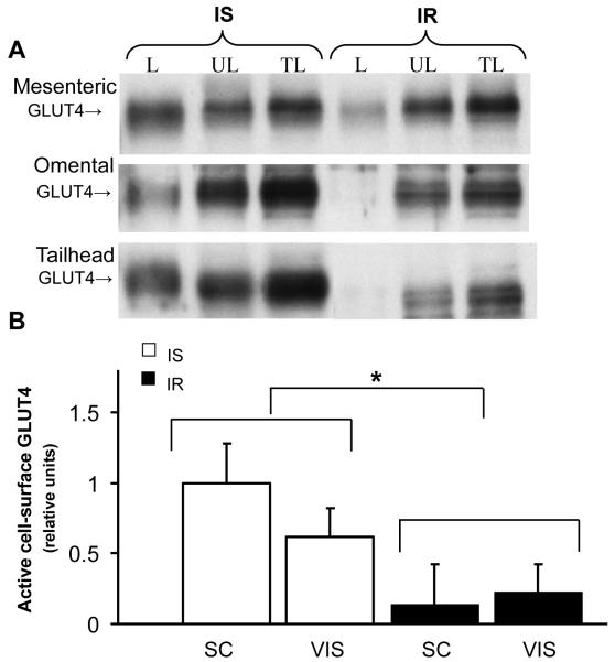
Figure 3.
Insulin resistance decreases active cell surface GLUT4 in photolabeled adipose tissue. (A): Representative Western blot of cell surface GLUT4 during insulin-sensitive (IS) and insulin resistant (IR) state; after cell-surface biotinylation of adipose tissue, streptavidin-isolated photolabeled GLUT4 was detected by immunoblotting. L: labeled fraction; UL: unlabeled fraction; TL: total lysate. (B) Mean ± SE of labeled cell-surface content of active GLUT4 in subcutaneous (Sc; tailhead) and visceral (Vis; omental and mesenteric) adipose sites during insulin-sensitive (IS) and insulin resistant (IR) state (n=2-4/group). Relative units were expressed in relation to an internal positive control; ‡: P<0.05. vs. IS group.

