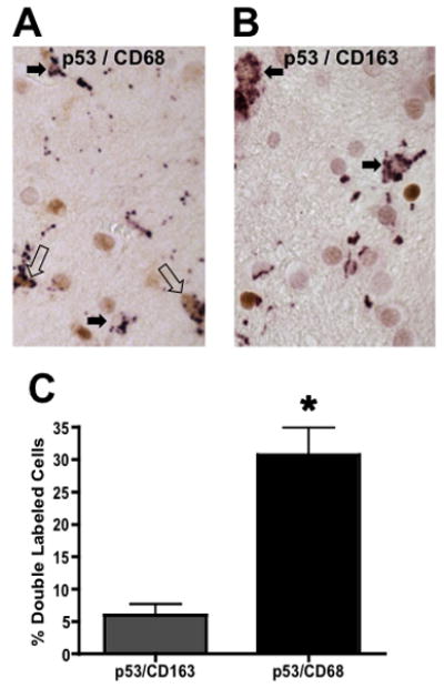
Figure 7.

Separate populations of microglia demonstrate immunoreactivity for a marker of alternatively activated and deactivated macrophages, CD163 or p53 activation in HAND cases. A) Human cortical tissue sections immunolabeled for p53 (brown) and CD68 (purple) reveal that a portion of microglia are immunoreactive for both markers. Open arrows point to double labeled cells and closed arrows identify CD68 single labeled cells. B) A section adjacent to the one in A is shown labeled with antibodies to p53 (brown) and CD163 (purple). Closed arrows point to CD163 labeled microglia. C) Quantification of double labeled microglia in adjacent sections from twelve HAND patients (*=p<0.0001 using a paired t-test).
