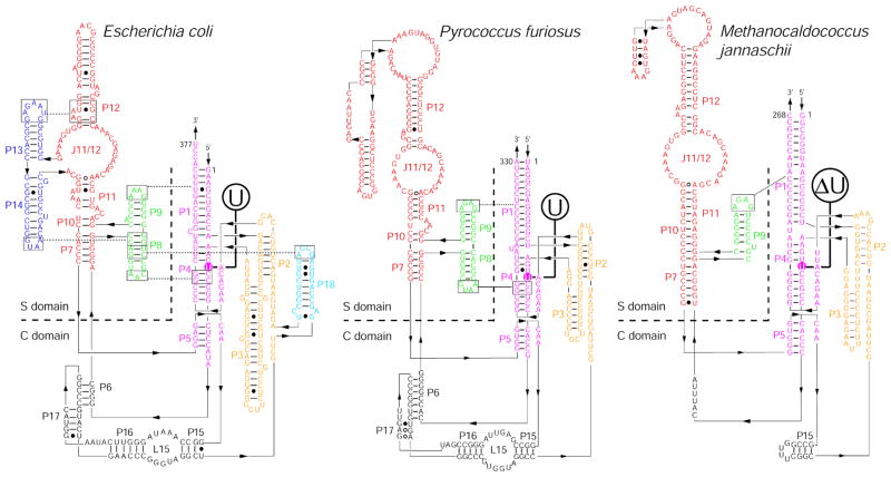
Figure 1.
Secondary structures of RPRs from Bacteria (e.g., Escherichia coli) and Archaea [e.g., Pyrococcus furiosus (Pfu; type A) and Methanocaldococcus jannaschii (Mja; type M)].27,75 The universally conserved bulged uridine in the P4 helix of all RPRs is enclosed in a colored circle. A deletion (ΔU) mutation made at this position in the Mja RPR is also indicated.

