Fig. 3.
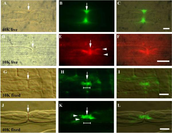
10kDa and 40kDa dextran tracer penetration in control sciatic nerve fibers. The node of Ranvier is marked by an arrow in A, D, G and J. Images A–F are from live injected fibers while G–L are from fixed fibers soaked in dextran solution.
A–C. 40kDa dextran (green). The tracer has pooled in the perinodal space on either side of the nodal slit (arrow, B). Scale bar = 5μm.
D–F. 10kDa dextran (red). The tracer has spread from the node of Ranvier (arrow, D, E) laterally through the paranode into the internode forming “hairpins” (arrowheads, E). Scale bar = 5μm.
G–I. 10kDa tracer (green) after 1hr soak showing the nodal slit (arrow G, H). Paranodal bar in H widens at its abnodal ends at the junction with the internode (shoulder). Scale bar = 10μm.
J–L. 40kDa tracer (green) showing paranodal bar and hairpins (arrowheads in K) after 2hr soak. The tracer is sometimes washed out of the node slit, which then appears dark (arrow, K). Scale bar =10μm.

