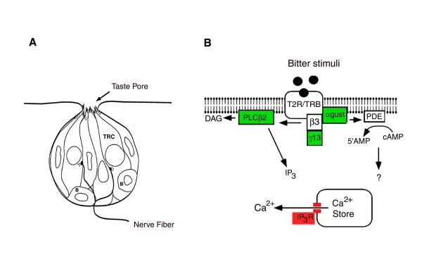
Figure 1.
Diagrammatic representation of a rodent taste bud and important components of the bitter transduction pathway. (A) A typical taste bud consists of 50-100 taste receptor cells (TRCs) that extend from the basal lamina to the taste pore. Taste stimuli interact with taste receptors on the apical membrane, while nerve fibers form chemical synapses with the basolateral membrane. Basal cells (labeled B) along the margin of the taste bud are proliferative cells that give rise to taste receptor cells. (B) Bitter stimuli interact with T2R/TRB receptors located on the apical membrane. These receptors couple to a heterotrimeric G protein consisting of α-gustducin, β3, and γ13. Alpha gustducin activates phosphodiesterase (PDE), causing decreases in intracellular cAMP, while β3γ13 activates phospholipase C β2 (PLCβ2) to produce the second messengers inositol 1,4,5 trisphosphate (IP3) and diacylglycerol (DAG). The IP3 binds to receptors located on smooth endoplasmic reticulum, causing a release of Ca2+ into the cytosol. The purpose of this study was to identify the IP3 receptor isotype that is expressed in taste cells.

