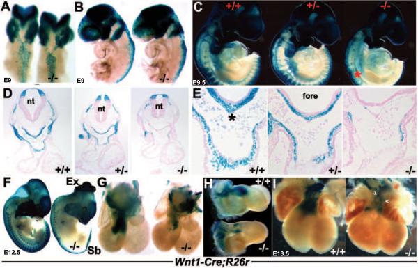
Figure 3. Wnt1Cre/R26R lineage mapping of NC cells and their derivatives.
(A,B) Dorsal (A) and left lateral (B) views of E9 wildtype and Pax3Δ5 null (−/−) X-Gal stained embryos, illustrating impaired Pax3Δ5 null NC emigration and migration towards the 2nd PA, when compared to wildtype littermates (embryos on left). However, 1st arch and cranial NC migration appears unaffected. (C) Right lateral views of E9.5 wildtype (+/+), heterozygous (+/ −) and Pax3 null (−/−) littermates showing fewer CNC populate the heterozygous 3/4/6th PAs (middle embryo), and still even less CNC colonize the Pax3 null 3/4/6th arches (right embryo). There is also a significant reduction in NC contribution to the Pax3 null dorsal root ganglia (indicated by * in C). However, cranial NC migration appears unaffected. (D,E) Histology through the OFT region of embryos shown in C, confirmed a lack of Wnt1Cre-marked CNC within the Pax3 null 4th PAs (D) and AP septum (indicated by * in wildtype in E) and OFT cushions. However, the dorsal-ventral boundary of Wnt1-Cre-marked neuroepithelial cell within the NT was similar amongst all three genotypes (D). (F) Lateral view of E12.5 wildtype and Pax3 null, illustrating extensive NC reduction along the anterior-posterior axis of the nulls (right embryo), absent lacZ staining of dorsal root ganglia, as well as spina bifida (Sb) and exencephaly (Ex). (G,H) Wholemount lacZ staining of Wnt1Cre reporter expression in E12.5 wildtype/Wnt1Cre/R26R and Pax3Δ5/Δ5/Wnt1Cre/R26R hearts. Note Pax3 null CNC reduced OFT colonization (−/−), viewed frontally (G) and from the right (), but that the length and width of the mutant OFT is similar to control. (I) Reduced CNC colonization is most evident in E13.5 Pax3 null hearts (right heart), and some lacZ-marked NC are ectopically located on the mutant OFT surface (arrowheads). Stage-matched embryos and isolated hearts are shown photographed at the same magnification.

