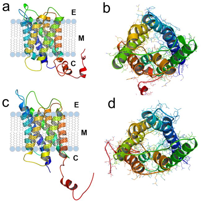
Fig. 1.
Three dimensional simulations of wild type mouse AQP0 (a) and AQP1 (b) proteins predicted using 3D-JIGSAW version 2.0. The cartoons were created using PyMOL. Monomers of mouse AQP0 and AQP1 are rendered in cartoon showing the folds, helix assignment, and location in the membrane. Modeled structures of mouse AQP0 and mouse AQP1 are in the same orientation. (a) and (c), ribbon diagrams of AQP0 and AQP1, respectively, with transmembrane helices embedded in the lipid bilayer. (b) and (d) AQP0 and AQP1, respectively; channels viewed across the cell membrane. In the middle, AQP0 water channel (b) is occupied by more side chains compared to AQP1 (d) that exhibits a wider aqueous pore.

