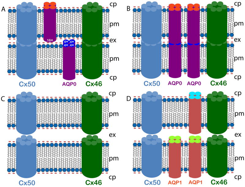
Fig. 6.
Schematic models illustrating AQP0 mediated fiber cell-to-fiber cell adhesion and cell-to-cell communication. Lens fiber cells express AQP0 in the membrane (~ 45%). AQP0 forms functional water channels as an analogous function. It also facilitates reduction in fiber cell extracellular space in the WT lenses in contrast to AQP0 knockout and TgAQP1+/+/AQP0−/− lenses, as shown by the EM studies (Fig. 5B), for reducing light scattering, and for increasing lens transparency and fiber cell communication through gap junction channels. AQP0 aids in pulling the fiber cell plasma membranes of opposing cells closer by functioning as an adhesion protein which is a unique role to maintain a streamlined architecture. Four models are proposed here, based on the available literature and our ultrastructural studies. Two possible working models for wild type mouse lens (A, B) show narrow extracellular space (see also Fig. 5B, c, d). A) Positively charged extracellular domain of AQP0 interacts with negatively charged lipid membrane to draw the membranes closer, and B) AQP0 extracellular domains of opposing fiber cell plasma membranes interact with each other to reduce the extracellular space. Two possible working models (C, D) show wide extracellular space. C) Wide extracellular space in the AQP0−/− (as seen in Fig. 5B, g, h) due to negatively charged fiber cell membranes opposing each other. D) Wide extracellular space between the TgAQP1+/+/AQP0−/− mouse lens fiber cells (see also Fig. 5B, k, l) due to the absence of a net +ve charge in the extracellular domains of AQP1, and/or due to the repulsion caused by negatively charged membranes facing each other. cp — cytoplasm, pm — plasma membrane, ex — extracellular space.

