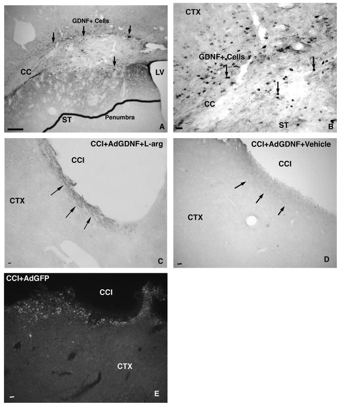
Figure 5.
GDNF expression –GDNF protein expression is robust in Sham animals one week post-injection, producing a penumbra of staining (outlined area in A) and significant numbers of GDNF+ Cells (B). Following injury, GDNF expression decreases significantly, with slightly more expression seen in the animals treated with AdGDNF+L-Arginine (C) versus AdGDNF alone (D). Following injury, GFP+ Cells are still present surrounding the contusion cavity (E). Scale bar - A,C, D, &E = 250μm; B=25 μm. Arrows point to GDNF penumbra and or GDNF+cells. LV=Lateral Ventricle; CC = Corpus Callosum; ST=Striatum; CTX =Cortex; CCI=Contusion cavity

