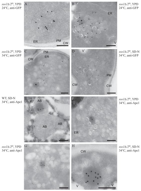
Figure 4. Ultrastructural Analysis of the Atg9-Containing Membranes and of the prApe1 Oligomer.
Wild-type (WT, UNY151, panel E) and sso1Δ/2ts (UNY141, panels A-D and F-H) cells were grown to exponential phase at PT (panels A and B) before being shifted to NPT (panels C-H) in either rich (YPD, panels A-C, F and G) or nitrogen starvation medium (SD-N, panels D, E and H) for 1.5 h. Cells were processed for immuno-EM as described in the Supplement before and after the temperature and the medium changes. Cryo-sections shown in panels A to D were immuno-labelled for GFP to detect Atg9-GFP, while those presented in panels E to H were immuno-labelled for Ape1. (A) A tubulovesicular cluster detected infrequently in the mutant strain.
(B) Overview of a cluster of secretory vesicles that also contain Atg9-positive carriers.
(C, D) The Atg9-containing vesicles are more dispersed throughout the cytoplasm in the sso1Δ/2ts mutant shifted to NPT.
(E) Autophagic bodies accumulated in the vacuole of nitrogen-deprived wild-type cells, and some of them contain the electron-dense prApe1 oligomer.
(F and G) The prApe1 oligomer is associated with vesicular structures in the sso1Δ/2ts mutant shifted to NPT in rich medium.
(H) In the sso1Δ/2ts cells nitrogen starved at NPT, the prApe1 oligomer is associated with 1-3 large vesicles. The asterisks mark the circular electron-dense structures that correspond to prApe1 oligomers, often observed in proximity to Atg9-positive membranes. Scale bar, 200 nm. AB, autophagic body; CW, cell wall; ER, endoplasmic reticulum; PM, plasma membrane; V, vacuole. See also Figure S4.

