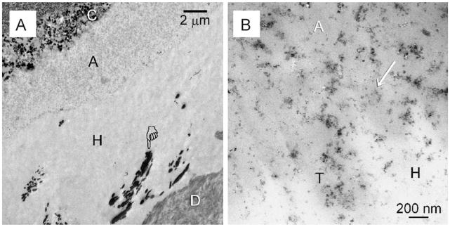
Fig. 1.
TEM images taken from unstained non-demineralised sections taken from the baseline control groups that were extracted immediately without ageing. This specimen was bonded using 2% chlorhexidine as a MMP inhibitor. The specimen was immersed in a silver nitrate tracer prior to TEM processing. (A) Silver-impregnated water-rich, resin-sparse regions (pointer) could be identified within the hybrid layer (H). C: composite; A: filled adhesive; D: dentine. Similar features were observed in the baseline control of premolars that were bonded without the use of chlorhexidine (not shown). (B) High magnification of (A). Nanofiller clusters (arrow) from the adhesive (A) could be seen in close proximity with the surface of the hybrid layer (H). T: dentinal tubule.

