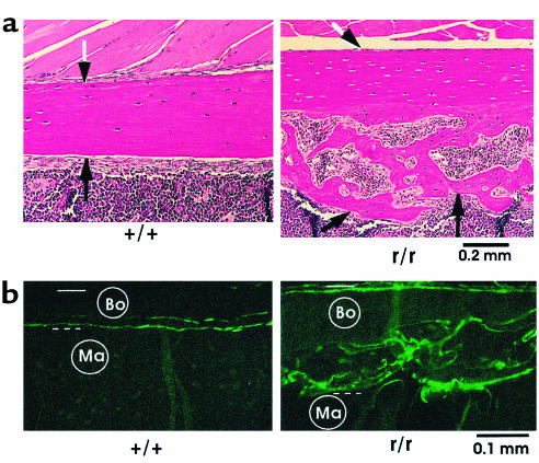
Figure 6.
Presence of excessive endosteal bone in femurs from Col1a1r/r mice. (a) Longitudinal histological sections stained with H&E of femurs from 10-month-old wild-type and Col1a1r/r mice. The location of the periosteal surface (black and white arrows) and endosteal surface (black arrows) is indicated. In wild-type mice the endosteal surface was smooth, and there was no evident endosteal new bone. In Col1a1r/r mice, in contrast, abundant endocortical new bone covered the endosteal surface. (b) Sections of femurs, embedded in methyl methacrylate and examined by fluorescence microscopy, from 10-month-old wild-type and Col1a1r/r mice injected twice with calcein, 30 days apart. Bo, bone; Ma, marrow area. The periosteal surface is indicated by the solid open bar and the endosteal surface by the dashed open bar. Note minimal calcein label at the endosteal surface of the femur from the wild-type mouse in contrast to the striking labeling of the new endosteal bone (a) in the femur from the Col1a1r/r mouse.

