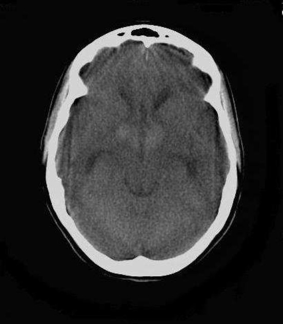
Figure 1.

Axial non enhanced CT (NECT) scan of brain (KV: 120, MAS: 60) of a 22-year-old woman with intrinsic third ventricular craniopharyngioma
At the level of temporal horns revealed mid line, homogenous, hyperdense mass below the bifrontal horns with mild obstructive hydrocephaly due to pressure effect on foramen of Monroe.
