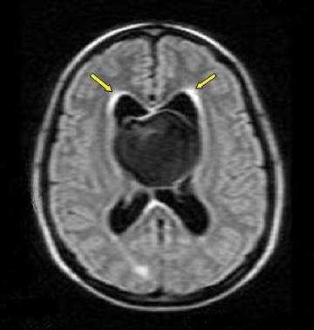
Figure 6.

Axial FLAIR image spin-echo (SE) of magnetic resonance imaging (MRI) of a 22-year-old woman with intrinsic third ventricular craniopharyngioma
The MRI of brain (philips intra 1.5 T, TE:140 msec, TR: 11000.6 msec) at the level of body lateral ventricles showed large midline hypointense mass, with periventricular hyperintensity due to interestial edema (arrow)
