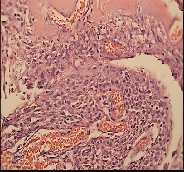
Figure 8.

Papillary craniopharyngioma in a 22-year-old woman patient On H&E staining (Magnification: ×40) it is microscopically composed of solid, well differentiated, pseudopapillary squamous epithelium with separation and desquamation of the epithelium.
