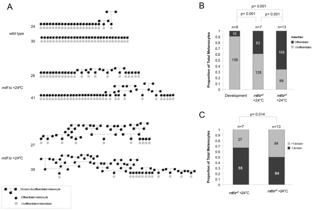
Fig. 5.
Hypomorphic Mitf activity enhances differentiated cell division. (A) Schematic representation of melanocyte development in embryos imaged by time-lapse microscopy. Two embryos are represented for each treatment condition. Wild-type or mitfavc7 mutant embryos were grown at 28.5°C for ∼20 hours, embedded in agarose, shifted to temperatures below 24°C (23-24°C) or over 24°C (25°C, 25.5°C or 26.0°C), and imaged by time-lapse microscopy until ∼108-151 hpf. All melanocytes begin as undifferentiated (grey) melanoblasts that become differentiated (black). Lineage is represented by broken lines: vertical broken lines indicate relative time between division events. The approximate order of melanocyte development is represented along the horizontal axis. Final total number of melanocytes in imaged region is indicated. (B) Stacked bar graph indicating the proportion of melanocytes that arise from differentiated or undifferentiated cells in wild type (n=9; 16/174 melanocytes) and mitf vc7 mutants grown at below 24°C (n=7; 82/210 total melanocytes) or over 24°C (n=13; 168/256 total melanocytes). The proportion of melanocytes arising from a differentiated cell is significantly greater in mitfvc7 mutants at below 24°C and over 24°C compared with wild-type fish; P<0.001; 95% CI (0.22, 0.38) and P<0.001; 95% CI (0.49, 0.64) respectively; binomial test of comparison of proportions. Additionally, the proportion of melanocytes arising from a differentiated cell between mitfvc7 mutants at below 24°C and over 24°C is significant; P<0.001; 95% CI (0.18, 0.36); binomial test of comparison of proportions. Datasets include only mitfavc7 time-lapse analysis that is longer than 108 hpf. (C) Bar graph indicating the proportion of mitfavc7 melanocytes that undergo serial division at below 24°C (n=7; 27/82) compared with those grown at over 24°C (n=13; 84/168). P=0.014, binomial test of comparison of proportions; 95% CI (0.04, 0.30). Datasets include only mitfavc7 time-lapse analyses that were longer than 108 hpf.

