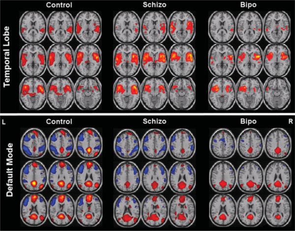
Fig. 3.
Used with permission from Calhoun et al. [18]. Two useful resting state networks: temporal lobe and default mode. These are group averages extracted from fMRI data from controls, schizophrenia patients, and bipolar patients, showing clear differences.

