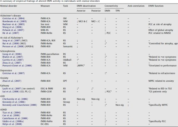
Fig. 6.
Used with permission from Broyd et al. [15]. Abbreviations and symbols used in this table: DMN, default-mode network; TPN, task-positive network; MRI, magnetic resonance imaging; fMRI, functional magnetic resonance imaging; EEG, electroencephalogram; ICA, independent component analysis; ROI, region-of-interest (or seed voxel); ReHo, regional homogeneity; NeHo, network homogeneity; FA, fractional anisotropy; RS, resting-state; SM, sensory motor task; EPT, emotional processing task; WM, working memory task; ADHD, attention-deficit/hyperactivity disorder; ASD, autism spectrum disorder; MCI, mild cognitive impairment; GS, generalized seizure patients; PS, partial seizure patients; C, controls; MMSE, mini mental state exam; (−) indicates that this was not tested; (↑) reflects an increase, for DMN deactivation it refers to increased deactivation; (↓) reflects a decrease, for DMN deactivation it refers to reduced deactivation; “non-sig” indicates the groups did not differ significantly; (*) indicates association between DMN function and particular result. All results are considered in comparison to a normal control group unless otherwise specified.

