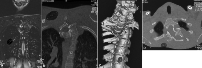
Fig. 2.
Case 1. Coronal MRI (A) and CT (B) images show two different planes, axial and coronal, of the dislocated spine in the same cut (the double-plane sign). Three-dimensional reconstruction of the CT scan shows complete dislocation of the spine (C). Axial cut of the CT scan shows the classic double-vertebrae sign of rotational dislocation of the spine.

