FIGURE 4.
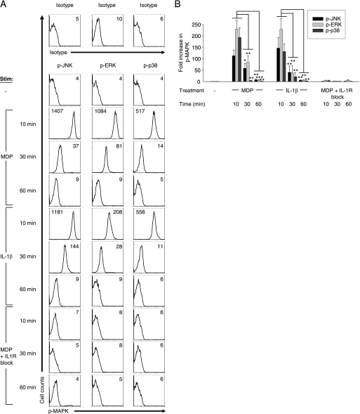
Decreased MAPK activation upon Nod2 signaling in the absence of the IL-1β autocrine loop persists over time. Human MDMs from healthy controls (n = 6–8) were stimulated with 100 μg/ml MDP, 10 ng/ml IL-1β, or 100 μg/ml MDP with 0.5 μg/ml IL-1Ra and 1 μg/ml anti-IL-1β antibody for 10, 30, or 60 min and analyzed by flow cytometry for the expression of phospho-JNK, phospho-ERK, or phospho-p38. A, shown are representative flow cytometry plots with the indicated mean fluorescence intensity values. Stimulated cells stained with isotype controls are shown. B, summarized data are represented as the -fold phospho-MAPK induction normalized to untreated cells + S.E. (error bars). *, p < 0.05; **, p < 0.01; ***, p < 0.001. p-, phospho-.

