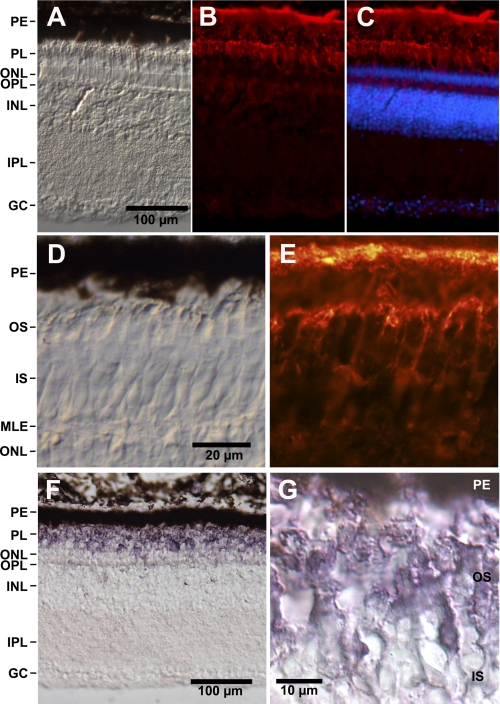
FIGURE 3.
Localization of GbE protein and mRNA in the chicken retina. A–C, longitudinal cryosections of the chicken retina. PE, pigment epithelium; PL, layer of outer and inner segments of photoreceptor cells; ONL, outer nuclear layer; OPL, outer plexiform layer; INL, inner nuclear layer; IPL, inner plexiform layer; GC, layer of ganglion cells. A, bright field microscopy image. Scale bar, 100 μm. B, indirect anti-GbE immunofluorescence. Bright immunofluorescence is visible in the pigment epithelium and the outer segments of photoreceptors; weak staining is visible in the outer plexiform layer and the ganglion cells. C, merged figure showing staining of the nuclei with Hoechst dye 33258. D and E, higher magnification of the photosensitive outer segments (OS) shows anti-GbE immunofluorescence of the cytoplasm surrounding the stacks of membrane-enclosed disks (E). D, bright field image. Scale bar, 20 μm. IS, inner segments; MLE, membrana limitans externa. F and G, immunohistological staining employing a secondary antibody coupled with alkaline phosphatase shows strong staining of the outer segments.

