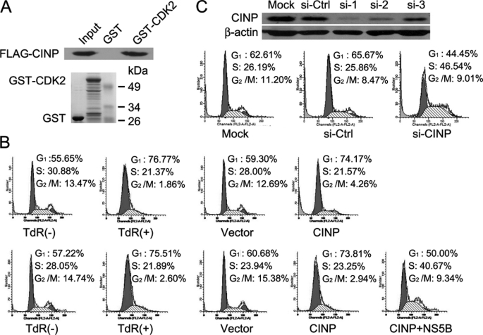
FIGURE 3.
Role of CINP in regulation of G1/S transition. A, the CDK2/CINP interaction was confirmed by a GST pulldown assay. B, HeLa cells were synchronized by the addition of 2 mm thymidine (TdR) for 16 h. After release, cells were transfected with CINP or empty vectors or co-transfected with CINP and NS5B. Forty-eight hours later, the cells were harvested and analyzed by flow cytometry. C, HeLa cells were transfected with 60 nm CINP-specific siRNA or control siGFP. Seventy-two hours later, the knockdown efficiency was detected by an anti-CINP antibody (upper panel). HeLa cells were then either mock-transfected or transfected with 60 nm siGFP or siCINP, respectively. Seventy-two hours post-transfection, the cells were analyzed for DNA content by flow cytometry. The results are representative of three independent experiments (lower panel). Ctrl, control.

