FIGURE 3.
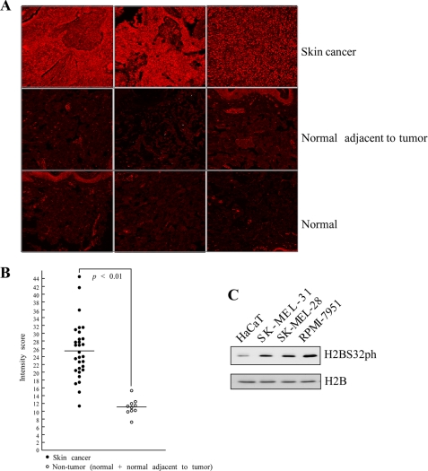
H2BS32ph is highly abundant in skin cancer tissues and skin cancer cell lines compared with normal counterparts. A, immunofluorescent staining and confocal microscopy were used to detect the levels of H2BS32ph in tissues from duplicate core samples of 9 cases of squamous cell carcinoma, 10 cases of basal cell carcinoma, 11 cases of malignant melanoma, and 5 each of adjacent normal tissue and normal tissue. H2BS32ph was detected by immunofluorescent staining with the H2BS32ph antibody as the primary antibody and Cy3-conjugated donkey anti-rabbit as the secondary antibody. Photographs from representative cases are shown. B, the intensity score of fluorescence from each sample was determined using Image J (National Institutes of Health); horizontal lines indicate the median fluorescence scores of skin cancer or noncancerous tissue samples; p < 0.01, Mann-Whitney U test. C, the level of H2BS32ph in several skin cell lines. Human keratinocytes (HaCaT) and several different human melanoma cell lines were screened by Western blot to determine the level of H2BS32ph. Total H2B protein was used to verify equal protein loading.

