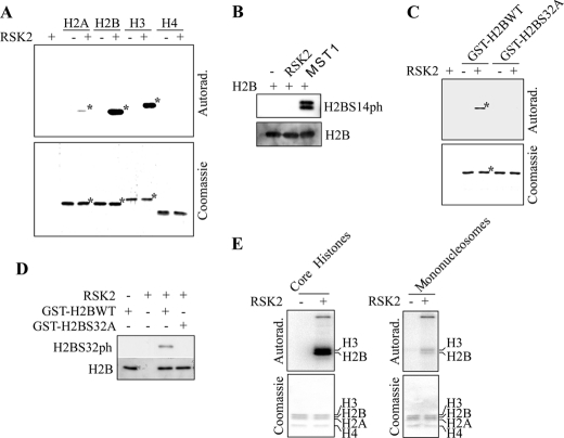
FIGURE 4.
RSK2 phosphorylates H2BS32. A, an in vitro kinase assay using active RSK2 and recombinant histone H2A, H2B, H3, or H4 as substrate in the presence of 32P was performed and results visualized by autoradiography (top panel). B, Western blot analysis of H2B phosphorylation after a kinase assay using active RSK2 or MST1 and recombinant H2B. A phospho-specific antibody was used to detect phosphorylation of H2BS14 (top panel). C, mapping of the H2B site phosphorylated by RSK2. A GST-H2B wild type or GST-H2BS32A mutant protein was used as substrate for active RSK2 in an in vitro kinase assay in the presence of 32P and visualized by autoradiography (top). D, Western blot analysis of H2B phosphorylation after a kinase assay using active RSK2 and GST-H2BWT or GST-H2BS32A mutant protein. The H2BS32ph antibody was used to detect phosphorylation of H2BS32 (top panel). For B and D, the same blot was also reprobed with an antibody against total H2B to monitor equal protein loading (bottom panels). E, RSK2 phosphorylates nucleosomal H2B. RSK2 phosphorylates H2B present in core histones (left panel) as well as H2B present as a component of the mononucleosomes (right panel). The experiments were performed as described under “Experimental Procedures,” except that different histone substrates were used for the kinase assays. Each asterisk indicates the respective protein band. For A, C, and E, the corresponding gels are stained with Coomassie Blue to monitor equal protein loading (bottom panels of each).

