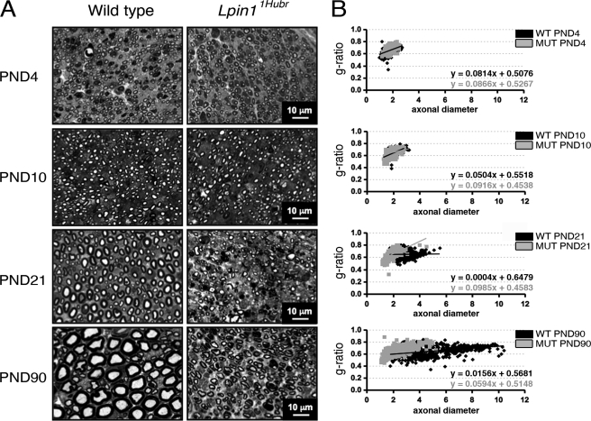
FIGURE 3.
Progression of myelination in Lpin11Hubr rats. A, toluidine blue-stained semi-thin sections from the medial region of the sciatic nerve of Lpin11Hubr and wild-type rats at PND 4, 10, 21, and 90. At PND 4 and 10, the level of myelination is similar in wild-type and mutant nerve. At PND 21 and 90, hypomyelination is visible in sciatic nerves of Lpin11Hubr rats compared with wild-type nerves. At PND 90, the general morphology of Lpin11Hubr sciatic nerves is improved as compared with the general morphology of sciatic nerves isolated from Lpin11Hubr rats at PND 21. B, at PND 4 and 10, average g-ratio, g-ratio trend line, and average axonal diameter of medial sciatic nerve tissue is equal between genotypes. At PND 21 and PND 90, axons of Lpin11Hubr rats (MUT) show a decreased axonal diameter as compared with wild-type (WT) axons, and an aberrant g-ratio trend line indicating hypomyelination. Axons of Lpin11Hubr rats, however, show an improvement in trend line at PND 90 as compared with PND 21, indicative of partial myelination recovery (PND 4, 10, 21, and 90; n = 2, n = 2, n = 2, and n = 1 per group, respectively; n = 107–374 axons per genotype).

