FIGURE 6.
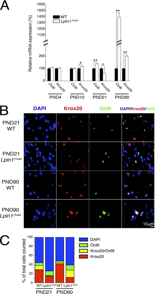
Myelination status of Schwann cells in Lpin11Hubr peripheral nerve. A, relative gene expression analysis of Oct6 and Krox20 (SC markers) in sciatic nerve tissue of Lpin11Hubr rats as compared with wild-type rats at PND 4, 10, 21, and 90 (n = 3, n = 3, n = 3, and n = 2 per group, respectively; *, p < 0.05; **, p < 0.001). B, sciatic nerve cross-sections of wild-type (WT) and Lpin11Hubr rats at PND 21 and 90 were immunostained with antibodies against transcription factors Krox20 (red) and Oct6 (green). The nuclei were stained with 4′,6-diamidino-2-phenylindole (DAPI; blue). C, the percentage of DAPI+ (blue), Krox20+ (red), Krox20+ and Oct6+ (yellow), and only Oct6+ (green) cells is shown (n = 2 per group). Data are expressed as mean ± S.E.

