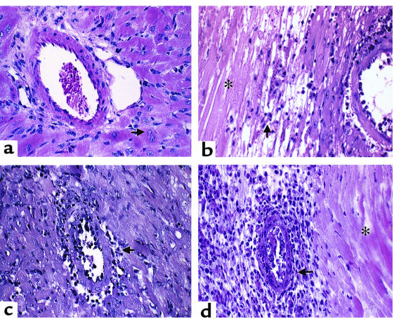
Figure 2.
Pathology of murine cardiac allograft rejection: syngeneic graft at day 8 after transplantation and allografts of WT, STAT4–/–, and STAT6–/– recipients. (a) Syngeneic graft: the artery shows no signs of inflammation and the cardiac muscle cells are intact, with viable nuclei (arrow). (b) Wild-type recipient graft: the small artery reveals inflammatory cells adherent to the endothelium. There is extensive infarction (coagulative or ischemic necrosis) of cardiac muscle cells (asterisk) and mononuclear inflammatory cell infiltration of the viable myocardium (arrow). (c) STAT4–/– recipient graft: there is active inflammation or endothelialitis of the inner layer of the small artery (arrow). The myocardium shows scattered lymphocytes. (d) STAT6–/– recipient graft: the active transmural inflammation (arrow) has resulted in extensive infarction of the myocardium (asterisk). There is also prominent inflammation of the parenchyma. Original magnification ×400 (H&E).

