FIGURE 6.
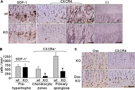
Decreased CXCR4 expression in epiphyseal growth plate of Osx::CXCR4fl/fl mice (KO) versus Cre-null littermate controls (WT). A, immunohistochemistry for SDF-1 expression in pre- and hypertrophic chondrocytes and CXCR4 expression in chondrocytic cells and in primary spongiosa (scale bar equals 50 μm). Negative (−) controls used isotype-matched control antibodies (scale bar equals 100 μm). B, histomorphometric analysis (OsteoII software, Bioquant) for the number of cells positive for SDF-1 or CXCR4 expression. The number of cells positive for SDF-1 immunostaining was measured from the pre- to hypertrophic zones. The number of cells positive for CXCR4 was measured in three regions (each at 185 × 250 μm2) in the middle of the growth plate or the primary spongiosa. Five measurements per section for three consecutive sections were taken to average each sample. C, immunohistochemistry for Osx and CXCR4 expression in growth plate of KO mice and control KO mice that were treated with doxycycline (200 μg/ml in drinking water) after birth (Dox-KO). n = 5 (three females and two males; 4 weeks old) WT or KO mice. Measurements are presented as mean ± S.D. *, p < 0.05 versus respective WT control.

