FIGURE 2.
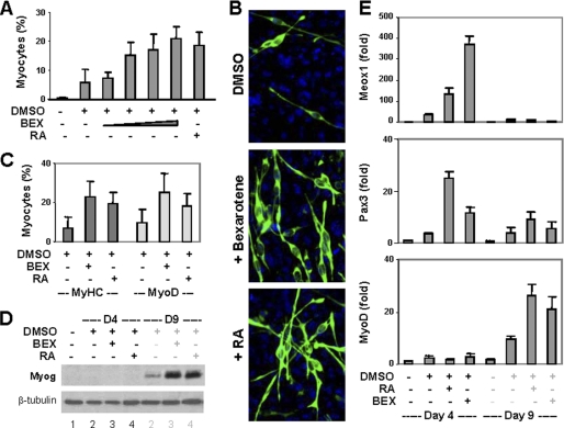
Effects of rexinoid on P19 cell differentiation. A, P19 aggregates were treated with bexarotene (BEX, 1 nm, 10 nm, 100 nm, or 1 μm) or RA (10 nm) in the presence of DMSO and stained for myosin heavy chain on day 9. Quantification is presented as fractions of cells differentiated into skeletal myocytes. Error bars are the S.D. of five independent experiments. B, the representative microscopic images. C, the cells were also costained for MyoD and quantified in comparison with myosin heavy chain (MyHC)-positive cells. D, Western blot analysis of myogenin expression with undifferentiated cells as control. β-tubulin was used as a loading control. E, the relative mRNA levels of Meox1, Pax3, and MyoD were determined by quantitative real-time RT-PCR and plotted as fold difference in relation to untreated day 4 controls after being normalized to GAPDH.

