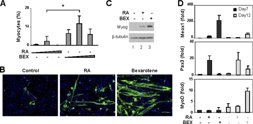
FIGURE 3.
Effects of rexinoid on ES cell differentiation. A, RA (5, 10, or 20 nm) or bexarotene (BEX, 20, 50, or 100 nm) were used during embryoid body formation. Cells were then plated on coverslips on day 7 and stained for myosin heavy chain and MyoD on day 20. Microscopic analyses were performed and plotted as fractions of cells differentiated into skeletal myocytes. *, p < 0.05. Error bars are the S.D. of four independent experiments. B, representative images of myosin heavy chain staining. C, Western analysis of myogenin with undifferentiated ES cells as control. β-tubulin was used as a loading control. D, the relative transcript levels of Meox1, Pax3, and MyoD were determined by real-time RT-PCR analysis and plotted as fold variance of untreated day 7 controls after being normalized to GAPDH.

