
FIGURE 2.
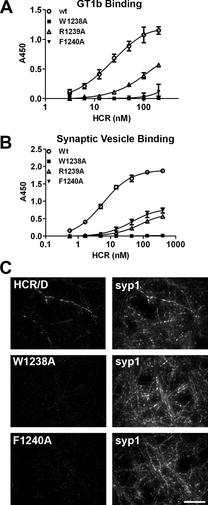
Role of the ganglioside binding loop in receptor recognition. A, GT1b (1 μg/well) was immobilized on a 96-well microtiter plate. HCR/D in indicated concentrations was bound for 1 h at 4 °C and detected as described in Fig. 1. HCR/D achieved 50% binding at ∼27 nm. B, isolated SVs were immobilized on a 96-well microtiter plate, and HCR affinity was determined as described under “Experimental Procedures.” HCR/D achieved 50% binding at ∼6 nm. C, HCRs were incubated with rat E18 primary cortical neurons for 5 min in depolarizing (56 mm potassium) or control (5.6 mm potassium) media. Cells were fixed and permeabilized with 0.1% Triton X-100 in 4% formaldehyde, blocked, and incubated with primary antibody overnight at 4 °C, and cells were then incubated with secondary antibody for 1 h at room temperature, fixed, and imaged. Representative images are shown; scale bar = 20 μm.