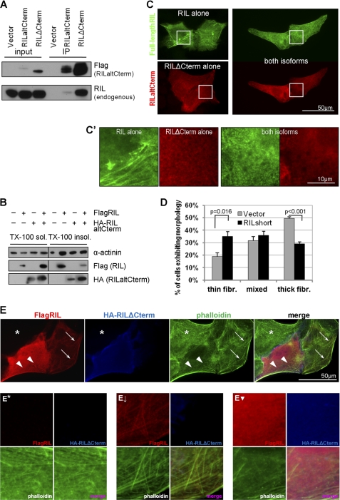
FIGURE 4.
RILaltCterm affects actin organization via interaction with full-length RIL. A, RILaltCterm binds to full-length RIL. Lysates from U2OS cells expressing FLAG-RILaltCterm were immunoprecipitated (IP) using anti-FLAG-agarose and probed with anti-RIL antibody. B, RILaltCterm changes distribution of full-length RIL between cytoskeleton-bound and cytosolic fractions and abrogates the effect of RIL on α-actinin-1. 293T cells were transfected as indicated and fractionated based on Triton X-100 solubility, and protein distribution was analyzed by immunoblotting. C, RILΔCterm affects the staining pattern of full-length RIL. U2OS cells were transfected with the indicated RIL constructs, fixed in 4% formaldehyde for 15 min, stained for FLAG and HA tags with suitable primary and fluorescently labeled secondary antibodies, and analyzed by wide field fluorescence microscopy. C′ shows enlarged detail of cell regions outlined in C. D, samples were processed as in C, and cells exhibiting thick fibers (fibr.), meshwork of thin fibers, or mixed staining pattern of full-length RIL in the presence or absence of RILaltCterm were counted. Bars represent an average of four independent experiments ± S.E., and p values were calculated by Student's t test. E, U2OS cells expressing FLAG-tagged full-length RIL and/or HA-tagged short isoform of RIL were grown on glass coverslips, fixed in 4% formaldehyde, and stained with rabbit anti-FLAG and rat anti-HA antibodies and then secondary AlexaFluor594 anti-rabbit and AlexaFluor350 anti-rat antibodies. F-actin was visualized by FITC-conjugated phalloidin. Asterisk indicates nontransfected cell; thick arrows indicate actin/RIL fibers; arrowheads indicate meshwork of thin actin fibers. E*, E↓, and E▾ represent enlarged detail of cells in E marked with asterisk, arrows, and arrowheads, respectively.

