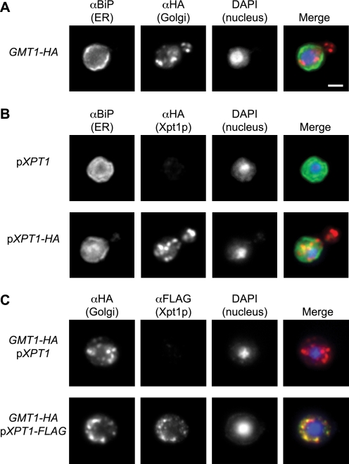
FIGURE 10.
Xpt1p-HA localization to the Golgi apparatus. Cryptococcal cells (JEC21) were fixed, permeabilized, immunolabeled, stained with DAPI, and visualized by immunofluorescence microscopy as detailed “Experimental Procedures.” Single channel and merged images are shown. Panel A, cells engineered to express an HA-tagged form of Gmt1p (a Golgi marker) were probed with antibodies to BiP (an ER marker) and HA, yielding distinct labeling patterns. Panel B, cells episomally expressing either wild-type or HA-tagged Xpt1p were probed with antibodies to BiP and HA, showing distinct labeling patterns. Panel C, Gmt1p-HA expressing cells (as in panel A) expressing either wild-type or FLAG-tagged Xpt1p were probed with antibodies to HA and FLAG, showing colocalization. All panels are at the same magnification; scale bar, 2 μm.

