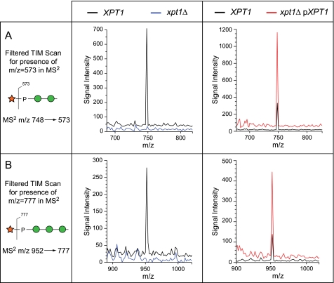
FIGURE 8.
Filtered TIM scans demonstrate Xyl-P-Man2 and Xyl-P-Man3 in O-linked glycans from wild-type, xpt1Δ mutant, and XPT1 overexpressing strains. Total ion mapping scans were filtered for the production of MS2 fragments that indicate the presence of Xyl-P motifs (panel A, m/z = 573 for Xyl-P-Man2; panel B, m/z = 777 for Xyl-P-Man3) in the indicated strains. MS2 is inherently more sensitive than full MS for ion-trap instruments, yet the Xyl-P modified glycans are detected as only minor deflections above base line in the xpt1Δ O-linked glycan preparations. The signal traces for KN99α in the left panels and for xpt1Δ pXPT1 in the right panels are displaced upward on the y axis by 5% of full scale for clarity.

