FIGURE 3.
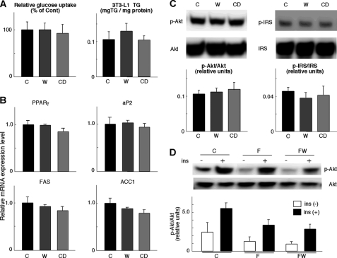
FXR agonists do not affect adipocyte differentiation and insulin sensitivity in vitro and in vivo. A, insulin-stimulated glucose uptake after adipocyte differentiation of 3T3-L1 cells with or without GW4064 or CDCA. Cells were stimulated with insulin (5 μg/ml) for 12 h, and the decrease of glucose in the medium was measured (left panel). TG content in these differentiated 3T3-L1 cells is shown in the right panel. C denotes control medium; W indicates medium with 10 μm GW4064; CD indicates medium with 20 μm CDCA. B, mRNA expression levels of Pparγ, aP2, Fas, and ACC1 in 3T3-L1 cells, as specified in A. C, determination of Akt/PKB or IRS-1 phosphorylation levels by Western blotting in differentiated 3T3-L1 cells. Cells were cultured with or without GW4064 or CDCA and stimulated with 5 μg/ml insulin for 5 min. Western blotting was performed with anti-Ser-473-phosphorylated Akt, total Akt, anti-Ser-307-phosphorylated IRS-1, or total IRS-1 antibodies. D, determination of Akt/PKB phosphorylation levels by Western blotting with anti-Ser-473-phosphorylated Akt or total Akt antibody in epWAT. Protein extracts were prepared from epWAT of C57BL/6J mice, which were fed a high fat diet with or without GW4064 for 76 days.

