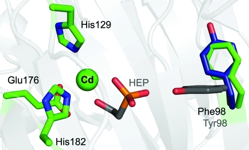
Figure 5.
Overlay of apo-HEPD, HEPD with 2-HEP bound, and HEPD-Y98F. Iron-binding residues, Cd(II), and Phe98 are colored green and are from the mutant structure (PDB entry 3RZZ). Colored gray are 2-HEP and Tyr98 from the liganded wt HEPD structure (PDB entry 3GBF). Colored blue is Tyr98 from apo-HEPD (PDB entry 3G7D). Tyr98 undergoes a torsional rotation into the active site as a result of a hydrogen bond that forms between the phenolic oxygen of tyrosine and a phosphonate oxygen of 2-HEP.

