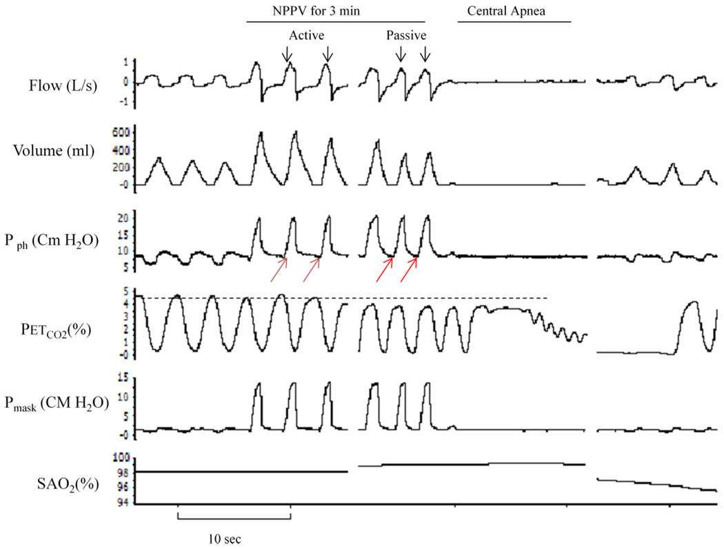
Figure 1.
Polygraph record of a trial that illustrates breaths during noninvasive positive pressure ventilation (NPPV) followed by central apnea. Ventilatory motor output inhibition was confirmed by the absence of negative pressure deflection on pharyngeal pressure (red arrows) and by the occurrence of central apnea upon termination of NPPV. First three breaths at the beginning of mechanical ventilation (active breaths) were compared to the last three breaths preceding the termination of NPPV (passive breaths). The horizontal dotted line indicates the level of end-tidal CO2 at baseline. Pph, pharyngeal pressure; PETCO2, end-tidal CO2; SaO2, oxygen saturation.

