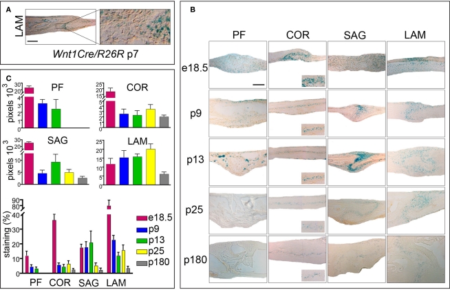
Figure 4.
Activation of canonical Wnt-signaling in embryonic and postnatal cranial sutures. (A) Xgal staining of LAM suture harvested from Wnt1Cre/R26R mouse. Xgal staining indicates the neural-crest origin of the LAM suture mesenchyme. The boxed area is enlarged to the right. (B) Xgal staining of PF, COR SAG, and LAM suture mesenchymes in Axin2+/− mice from e18.5 to p180. Scale bar: 100 μm. Inserts represent zoomed areas of the coronal sutures. Dashed lines indicate the bone plates. (C) Quantification of Xgal staining of different cranial suture mesenchymes in Axin2+/− mice at day p25 and p180.

